script async src="https://pagead2.googlesyndication.com/pagead/js/adsbygoogle.js">

Join On Whatsapp for Updates

Join On Whatsapp for Updates
Click to join whatsapp Group

Follow us

Showing posts with label Articles. Show all posts

Monday, April 13, 2020

People power: How India is attempting to slow the coronavirus


A policeman walks past barricades in front of the Vikasa Soudha building, Bengaluru, India.
India has been in lockdown because of the coronavirus pandemic since 25 March.Credit: Manjunath Kiran/AFP/Getty
Like many nations, India does not have enough kits to test most of its population for the new coronavirus. The country is instead relying on people power: thousands of health-care workers are fanning out across the country to trace and quarantine people who might have had contact with those with COVID-19. People are typically only tested if they develop symptoms.
Countries such as South Korea isolated infected people based on widespread testing, but some scientists say that India’s mass-surveillance approach could achieve a similar goal, and be relevant for other low- and middle-income countries facing kit shortages.

For Details Click--
 Nature, Gayathri Vaidyanathan,doi: 10.1038/d41586-020-01058-5

How does COVID-19 kill? Uncertainty is hampering doctors’ ability to choose treatments


How does COVID-19 kill? Uncertainty over whether it is the virus itself — or the response by a person’s immune system — that ultimately overwhelms a patient’s organs, is making it difficult for doctors to determine the best way to treat patients who are critically ill with the coronavirus.

Clinical data suggest that the immune system plays a part in the decline and death of people infected with the new coronavirus, and this has spurred a push for treatments such as steroids that rein in that immune response. But some of these treatments act broadly to suppress the immune system, stoking fears that they could actually hamper the body’s ability to keep the viral infection in check.

For Details read--
Nature Magazine
doi: 10.1038/d41586-020-01056-7

Structure of the RNA-dependent RNA polymerase from COVID-19 virus


A novel coronavirus (COVID-19 virus) outbreak has caused a global pandemic resulting in tens of thousands of infections and thousands of deaths worldwide. The RNA-dependent RNA polymerase (RdRp, also named nsp12) is the central component of coronaviral replication/transcription machinery and appears to be a primary target for the antiviral drug, remdesivir. We report the cryo-EM structure of COVID-19 virus full-length nsp12 in complex with cofactors nsp7 and nsp8 at 2.9-Å resolution. In addition to the conserved architecture of the polymerase core of the viral polymerase family, nsp12 possesses a newly identified β-hairpin domain at its N terminus. A comparative analysis model shows how remdesivir binds to this polymerase. The structure provides a basis for the design of new antiviral therapeutics targeting viral RdRp.

For More Details Visit and Click

How does a Coronavirus Test Work?


The most widely used test uses polymerase chain reaction or PCR, a technique invented in the 1980s. Central to PCR is its ability to “amplify” DNA — multiply genetic material into large enough quantities to analyse. In the case of the coronavirus, the virus’ RNA first needs to be converted to DNA using an enzyme called reversetranscriptase before putting samples into a PCR machine. Samples collected in throat and nasal swabs are mixed with reagents, which are particles that bind to the virus’s genetic material to ensure no other type of DNA in the sample is amplified. Next, the sample is placed in the PCR machine, which uses cycles of heating and cooling to help the reagents amplify the target DNA into millions of copies. A fluorescent dye is added — the dye glows if the result is positive.

For Details Click

Mutant enzyme could vastly improve recycling of plastic bottles


Recycling isn’t as guilt-free as it seems. Only about 30% of the plastic that goes into soda bottles gets turned into new plastic, and it often ends up as a lower strength version. Now, researchers report they’ve engineered an enzyme that can convert 90% of that same plastic back to its pristine starting materials. Work is underway to scale up the technology and open a demonstration plant next year.
“This is a huge step forward,” says John McGeehan, who directs the center for enzyme innovation at the University of Portsmouth and who was not involved with the work.
Polyethylene terephthalate (PET) is one of the world’s most commonly used plastics, with some 70 million tons produced annually. PET bottles are already recycled in many places. But the current approach has problems. For starters, recycling companies typically end up with a broad mix of different colors of the plastic. They then use high temperatures to melt those down, producing a gray or black plastic starting material that few companies want to use to package their products.
For More Details Click

Wednesday, November 6, 2019

NASA Guide to Air-filtering Houseplants

Sunday, October 27, 2019

Happy Diwali to all


Thursday, October 24, 2019

Genome and Epigenome

By Ms. Rabia.
B.Sc. Biotechnology- I
Meerut College Meerut

Wednesday, October 16, 2019

ICAR develops diagnostic kits for Japanese encephalitis

15th October, 2019, New Delhi
Dr. Trilochan Mohapatra, Secretary (DARE) & DG (ICAR) and Shri Atul Chaturvedi, Secretary, Department of Animal Husbandry, Government of India released 2 Diagnostic ELISA Kits for the control of Swine and Detection of Antigen at the Krishi Bhawan, New Delhi here today.
Release of Diagnostics Kits developed by ICAR-IVRI
In his address, Dr. Trilochan Mohapatra briefed about the main characteristics of the two Kits. Highlighting about the Japanese Encephalitis (JE) ELISA Kit (IgM) for Swine, Dr. Mohapatra mentioned that the JE is a re-emerging viral zoonotic disease leading to death of children every year in the country.
The kit developed by the ICAR-Indian Veterinary Research Institute is really helpful for assessing the active infection of JE virus in the swine population which predicts the outbreak of JE in the humans. As compared to the commercial kit available in the market at a price of Rs. 52,000; the ICAR-IVRI developed is available for the farmers at a minimal price of Rs. 5,000 only. The Director General mentioned that each kit is meant for testing around 45 samples.
Accentuating on the features of another Kit entitled “Bluetongue: Sandwich ELISA for detection of Antigen”, Dr. Mohapatra stated that the Bluetongue (BT) virus is an insect-transmitted viral disease of domestic and wild ruminants that includes the camelid species. The disease is widespread among the sheep, goats, cattle, buffaloes and camels in the country.
With the help of the Kit, the Bluetongue Virus can be controlled with the vaccination of susceptible animals, vector control and quarantine of infected animals with the good management practices. Apart from the vaccination, the early diagnosis and isolation of the infected animals are one of the commonly suggested preventive methods for controlling the spreading of the disease.
Dr. Mohapatra stated that the Kits have been validated earlier by the scientists for their credibility. The Kits have been prepared under the “Make in India” initiative of the Hon’ble Prime Minister, Shri Narendra Modi.
Shri Atul Chaturvedi remarked the joint initiative by the ICAR and the Department of Animal Husbandry as an incredible initiative. Shri Chaturvedi stated that the lunching of these two Kits will definitely be beneficial for not only the farming community, but the society as a whole. Citing about the number of casualties caused by the deadliest diseases in the societies every year, the Secretary mentioned that the Kits will prove to be a real helping hand. He also accentuated that the initiative will play a pivotal role in realizing the Hon’ble Prime Minister’s aim of doubling the farmer’s income.
Shri Sushil Kumar, Additional Secretary (DARE) & Secretary (ICAR) applauded the ICAR & Department of Animal Husbandry’s initiative in the designated direction. Shri Kumar stated that the kits have been prepared with care. He accentuated that proper research works by the scientists of both the organizations were carried out before the official launching of the Kits. The Kits were also tested against the mentioned virus and diseases caused by the animals for their validity.
Dr. Praveen Malik, Commissioner, Department of Animal Husbandry stressed on the prevention of the Bluetongue Virus that is affecting the animal’s health on a larger scale. The indigenously prepared Kit will definitely be helpful in fighting the infections caused by the virus effectively. The Kit has a tremendous potential for the detection of the virus.
Dr. R.K. Singh, Director, Indian Veterinary Research Institute emphasized on the measures to be adopted for controlling the spread of various harmful diseases by the birds, pigs and other animals. He stressed that the humans are more prone to the transmittable diseases. He expressed his concerns about the various cases in which the children are more affected by the diseases as compared to the younger or older ones. He mentioned that the Kits will provide help in achieving our targets effectively and efficiently.
Dr. Ashok Kumar, ADG, Animal Husbandry, ICAR along with other dignitaries of both the organizations were present during the occasion.
(Source: ICAR-Directorate of Knowledge Management in Agriculture, New Delhi)

Saturday, October 12, 2019

पत्तियों के रंग के अनुसार पोषक तत्वों की कमी की जानकारी

Information by- UP Agriculture Department

Color of Leaves tells. about the deficiency of particular mineral in plant.

for more details click

स्वच्छ पर्यावरण का स्वास्थ्य पर प्रभाव- जागरूकता अभियान


मेरठ के भारतीय गर्ल्स इंटर कॉलेज में पर्यावरण के संबंध में एक जागरूकता अभियान 11/10/2019  को आयोजित किया गया।
इस अभियान को विज्ञान प्रौद्यगिकी परिषद, उत्तर प्रदेश के सौजन्य से जिला विज्ञान कॉर्डिनेटर श्री दीपक शर्मा जी ने करवाया एवं शहर के गणमान्य विद्वानों ने भी अपना योगदान दिया। विज्ञान प्रसार के लिए अतुलनीय योगदान करने को 'आओ विज्ञान करके देखे'- youtube channel का धन्यवाद।


Friday, October 11, 2019

Indian Scientist share the Magic of Li-ion battery

John Goodenough, M. Stanley Whittingham and Akira Yoshino have been awarded the Noble Prize in Chemistry for "creating the rechargeable world" by developing lithium-ion batteries. Interestingly, an Indian scientist Samar Basu, played a crucial role in the development of viable lithium-ion batteries. After retirement, he returned from the US to India and motivated research on Lithium batteries in Indian institutions.
Storing electricity
Otto von Guericke's static electric generator and Michael Faraday's dynamo showed how electricity can be generated. The generated electrical energy had to be transmitted through a wire and consumed as soon as it was produced. Until Alessandro Volta invented the battery, there was no way of storing or transporting it.
There are three essential elements in any battery - anode, the negative end of the battery; cathode the positive end of the cell; and electrolyte a gel-like substance with chemical energy. Apart from this, some have a fourth component, a separator that keeps the anode and cathode apart to prevent short circuit. The electrical power in the battery is stored in the form of chemical energy and released when the electrochemical reaction takes place.
The electrolyte liquid or paste-like substance contains electrically charged particles or ions. When in contact with the anode, the electrolyte undergoes oxidation reaction. Two or more ions combine with the anode to form a compound, and one or more electrons are released. Simultaneously, the cathode undergoes a reduction reaction with the electrolyte. Ions and free electrons combine with cathode and form compounds.
During the oxidation-reduction (redox) electrochemical reaction, free electrons congregate around the anode. As a result, the anode and cathode are negatively and positively charged, respectively. A potential difference between the two ends is generated. The electrons from the anode are itching to move towards the cathode. The separator keeps the electrons at bay, and the reaction is under stalemate.
However, once you place this battery, in a flashlight and flip the switch on, a new pathway between the positive and the negative terminal of the battery is established. The electrical charge moves through the wire, from one terminal to the other in the cell completing the circuit. On its way around, the current passes through the filament in the bulb. The resistance of the filament makes it heat up and radiate heat and light. Once the circuit is complete, the redox reaction continues to take place until the electrodes run out of reagents for their respective reactions. Once the stored chemical energy is used up, the electric current stops and the battery is 'dead'.
Rechargeable batteries
Unlike ‘use and throw’ batteries used in a flashlight, typically the battery used in an automobile is rechargeable. The rechargeable batteries have unique materials as anode, cathode and electrolyte. When you plug such rechargeable battery into a power source, electrical current supplies electrons to the anode. Further, the electrons from the cathode are removed. The reverse chemical reaction restores the anode, cathode and the electrolyte to the near-original state, which we call as researching. Recharge is reverse of discharge of a battery.
One of the very widely used rechargeable batteries is lead-acid battery. In this battery the negative and positive plates are made of lead and lead dioxide respectively. The electrolyte, sulphuric acid, reacts with the plates to form lead sulfate. As more lead sulfate is produced, the charge in the battery goes down. When the battery connected to the power supply in the reverse direction, lead sulfate reverts to lead, lead dioxide and sulphuric acid and once again it is recharged.
Dynamos, dry cells, rechargeable lead-acid batteries were all adequate for the Industrial Revolution until the 1950s until the emergence of the semiconductor electronic devices. Electrical power was used typically in motor, electromagnet and vintage radio receivers made with bulky valves. The development of electronic devices required electrical power devices that are compact, potent and durable.
‘Use and throw’ zinc carbon battery will do for a flashlight. The rechargeable lead-acid batteries are excellent, to give a punch of energy at the turn of the ignition key to kick off the starter motor and crank the engine spring to life.


There are three essential elements in any battery - anode, the negative end of the battery; cathode the positive end of the cell; and electrolyte a gel-like substance with chemical energy.
But think of a battery sitting inside a pacemaker, prodding the heart to tick. You don't want that to stop forever. Nor the battery unit can be bulky. Consumer electronics devices such as electronic watches, toys, cameras, mobile phones and laptops also require robust, enduring batteries that pack more power in lightweight package.
Distinct chemistry of various types of batteries results in voltage output ranging from 1.0 to 3.6 V. By serially stacking cells, voltage can be multiplied and by parallel connection current can be increased. By suitable combination, we can get the desired output. The problem was to find a battery that is light in weight, yet gives more punch of energy per kilogram of mass.
Whittingham, Goodenough and Yoshino found the way. They share this year's Noble chemistry prize for this radical discovery that made the mobile revolution possible. Compared to the energy density of 0.13 of zinc copper flashlight batteries, and the 0.14 of lead acid batteries, the lithium-ion batteries have a density of 0.70 Mj/Kg. While the lead-acid batteries can be recharged typically 500 times, the lithium ion batteries can be cycled 500–1000 times.
Lithium magic
With just three electrons and three protons, lithium is the third lightest of all elements. With two of the three electrons making a pair, lithium happily lets the third one wander away as a free electron. What’s more, the electron peels off easily compared to other elements. The energy needed to knock off one electron of lithium is almost half that of Zinc or Cadmium, other typical anode metals. Lithium-ion can store about 10 times as much energy as lead-acid or 5 times as much as nickel-cadmium. It is an excellent material for battery, but for the fact that it is dangerously reactive. Pure lithium burst into flames when it comes in contact with water.
M. Stanley Whittingham began experimenting with lithium as an anode material during the 1970s. Along with lithium anode, he used titanium disulfide as the cathode. In the discharge phase, when the battery was connected to a device, the lithium atom released an electron to become an ion. The positive lithium-ion moved towards the cathode. Titanium disulfide has a lattice structure and the ions snuggled between the layers. The circuit was completed, and the battery produced a 2 volts current. When the battery was recharged, the lithium ions flew back across the electrolyte to their starting position at the anode. Cathode and anode returned to its original state.
But there were two challenges. As lithium reacted violently, the anode had to be isolated from water and air. The electrolyte had to be a non-aqueous solution. Whittingham was able to identify a suitable organic electrolyte from other researches to overcome this hurdle. But the second one was serious. As the battery discharged and recharged, lithium crystals grow into a wispy, needle-like structure known as dendrites connecting anode and cathode. This was disastrous. Once such a defect forms, the battery short-circuited and at times even exploded.
Meanwhile, John Goodenough at Oxford was studying properties of metal oxides. He realized that a metal oxide can soak up more electrons than metal sulfide. He found that the cobalt oxide and the titanium disulfide both had similar lattice structure. Goodenough figured that like the titanium disulfide, cobalt oxide can also capture lithium ions during battery discharge and release it during recharge. In addition, cobalt oxide could house more ions than titanium disulfide. Energy potential doubled with this swap. Goodenough's design generated 4 volts, double that of Whittingham. Yet the problem of naked lithium remained.
Meanwhile, Samar Basu at Bell Labs in the US showed that lithium ions could embed in graphite. He developed a new battery with niobium selenide as cathode host and graphite as the anode host. The electrolyte was salt of lithium dissolved in an organic solvent. Both the anode and cathode could implant lithium-ion. Once the external circuit was switched on, the lithium ions were drawn from the graphite towards the niobium selenide, and the free electrons moved in the reverse direction. During the charging, the electrons could push the lithium ions back to graphite host. This was the first lithium-ion rechargeable battery where the lithium ions swung back and forth between anode and cathode during discharge and charge. As there was no free lithium, the battery was safer.
The next big step came when Akira Yoshino tried to use petroleum coke, a by-product of oil production, as an anode. The layers of carbon in petroleum coke could soak up lithium ions efficiently when charged. Goodenough's metal oxide cathode, Yoshino's carbon layer anode were combined to produce yet another version of the lithium-ion batteries. In the absence of pure lithium, the concerns of safety and dendrites formation vanished. The voltage was still just 4, but the new cocktail was safe, durable, lightweight and rechargeable. It could withstand hundreds of cycling. The lithium-ion battery technology matured. The new batteries hit the market around 1991.
India Science Wire
[Images ©Johan Jarnestad/The Royal Swedish Academy of Sciences]

CRISPR/Cas9 & Targeted Genome Editing

What is CRISPR/Cas9?

The functions of CRISPR (Clustered Regularly Interspaced Short Palindromic Repeats) and CRISPR-associated (Cas) genes are essential in adaptive immunity in select bacteria and archaea, enabling the organisms to respond to and eliminate invading genetic material. These repeats were initially discovered in the 1980s in E. coli (9), but their function wasn’t confirmed until 2007 by Barrangou and colleagues, who demonstrated that S. thermophilus can acquire resistance against a bacteriophage by integrating a genome fragment of an infectious virus into its CRISPR locus (10).
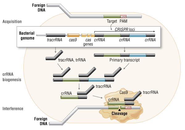
Three types of CRISPR mechanisms have been identified, of which type II is the most studied. In this case, invading DNA from viruses or plasmids is cut into small fragments and incorporated into a CRISPR locus amidst a series of short repeats (around 20 bps). The loci are transcribed, and transcripts are then processed to generate small RNAs (crRNA – CRISPR RNA), which are used to guide effector endonucleases that target invading DNA based on sequence complementarity (Figure 1) (11).

Figure 1. Cas9 in vivo: Bacterial Adaptive Immunity

Cas9 in vivo: Bacterial Adaptive Immunity

Cas9 in vivo: Bacterial Adaptive Immunity

In the acquisition phase, foreign DNA is incorporated into the bacterial genome at the CRISPR loci. CRISPR loci is then transcribed and processed into crRNA during crRNA biogenesis. During interference, Cas9 endonuclease complexed with a crRNA and separate tracrRNA cleaves foreign DNA containing a 20-nucleotide crRNA complementary sequence adjacent to the PAM sequence. 
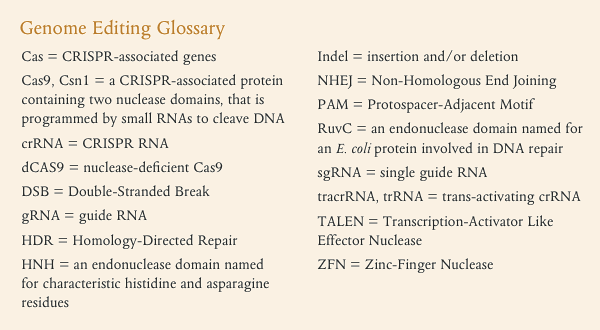
Genome Editing Glossary
One Cas protein, Cas9 (also known as Csn1), has been shown, through knockdown and rescue experiments to be a key player in certain CRISPR mechanisms (specifically type II CRISPR systems). The type II CRISPR mechanism is unique compared to other CRISPR systems, as only one Cas protein (Cas9) is required for gene silencing (12). In type II systems, Cas9 participates in the processing of crRNAs (12), and is responsible for the destruction of the target DNA (11). Cas9’s function in both of these steps relies on the presence of two nuclease domains, a RuvC-like nuclease domain located at the amino terminus and a HNH-like nuclease domain that resides in the mid-region of the protein (13).
To achieve site-specific DNA recognition and cleavage, Cas9 must be complexed with both a crRNA and a separate trans-activating crRNA (tracrRNA or trRNA), that is partially complementary to the crRNA (11). The tracrRNA is required for crRNA maturation from a primary transcript encoding multiple pre-crRNAs. This occurs in the presence of RNase III and Cas9 (12).
During the destruction of target DNA, the HNH and RuvC-like nuclease domains cut both DNA strands, generating double-stranded breaks (DSBs) at sites defined by a 20-nucleotide target sequence within an associated crRNA transcript (11, 14). The HNH domain cleaves the complementary strand, while the RuvC domain cleaves the noncomplementary strand.
The double-stranded endonuclease activity of Cas9 also requires that a short conserved sequence, (2–5 nts) known as protospacer-associated motif (PAM), follows immediately 3´- of the crRNA complementary sequence (15). In fact, even fully complementary sequences are ignored by Cas9-RNA in the absence of a PAM sequence (16).

Cas9 and CRISPR as a New Tool in Molecular Biology

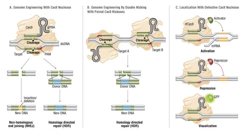
The simplicity of the type II CRISPR nuclease, with only three required components (Cas9 along with the crRNA and trRNA) makes this system amenable to adaptation for genome editing. This potential was realized in 2012 by the Doudna and Charpentier labs (11). Based on the type II CRISPR system described previously, the authors developed a simplified two-component system by combining trRNA and crRNA into a single synthetic single guide RNA (sgRNA). sgRNAprogrammed Cas9 was shown to be as effective as Cas9 programmed with separate trRNA and crRNA in guiding targeted gene alterations (Figure 2A).
To date, three different variants of the Cas9 nuclease have been adopted in genome-editing protocols. The first is wild-type Cas9, which can site-specifically cleave double-stranded DNA, resulting in the activation of the doublestrand break (DSB) repair machinery. DSBs can be repaired by the cellular Non-Homologous End Joining (NHEJ) pathway (17), resulting in insertions and/or deletions (indels) which disrupt the targeted locus. Alternatively, if a donor template with homology to the targeted locus is supplied, the DSB may be repaired by the homology-directed repair (HDR) pathway allowing for precise replacement mutations to be made (Figure 2A) (17, 18).
Cong and colleagues (1) took the Cas9 system a step further towards increased precision by developing a mutant form, known as Cas9D10A, with only nickase activity. This means it cleaves only one DNA strand, and does not activate NHEJ. Instead, when provided with a homologous repair template, DNA repairs are conducted via the high-fidelity HDR pathway only, resulting in reduced indel mutations (1, 11, 19). Cas9D10A is even more appealing in terms of target specificity when loci are targeted by paired Cas9 complexes designed to generate adjacent DNA nicks (20) (see further details about “paired nickases” in Figure 2B).
The third variant is a nuclease-deficient Cas9 (dCas9, Figure 2C) (21). Mutations H840A in the HNH domain and D10A in the RuvC domain inactivate cleavage activity, but do not prevent DNA binding (11, 22). Therefore, this variant can be used to sequence-specifically target any region of the genome without cleavage. Instead, by fusing with various effector domains, dCas9 can be used either as a gene silencing or activation tool (21, 23–26). Furthermore, it can be used as a visualization tool. For instance, Chen and colleagues used dCas9 fused to Enhanced Green Fluorescent Protein (EGFP) to visualize repetitive DNA sequences with a single sgRNA or nonrepetitive loci using multiple sgRNAs (27).

Figure 2. CRISPR/Cas9 System Applications

CRISPR/Cas9 System Applications

A. Wild-type Cas9 nuclease site specifically cleaves double-stranded DNA activating double-strand break repair machinery. In the absence of a homologous repair template non-homologous end joining can result in indels disrupting the target sequence. Alternatively, precise mutations and knock-ins can be made by providing a homologous repair template and exploiting the homology directed repair pathway.
B. Mutated Cas9 makes a site specific single-strand nick. Two sgRNA can be used to introduce a staggered double-stranded break which can then undergo homology directed repair.
C. Nuclease-deficient Cas9 can be fused with various effector domains allowing specific localization. For example, transcriptional activators, repressors, and fluorescent proteins.

Targeting Efficiency and Off-target Mutations

Targeting efficiency, or the percentage of desired mutation achieved, is one of the most important parameters by which to assess a genome-editing tool. The targeting efficiency of Cas9 compares favorably with more established methods, such as TALENs or ZFNs (8). For example, in human cells, custom-designed ZFNs and TALENs could only achieve efficiencies ranging from 1% to 50% (29–31). In contrast, the Cas9 system has been reported to have efficiencies up to >70% in zebrafish (32) and plants (33), and ranging from 2–5% in induced pluripotent stem cells (34). In addition, Zhou and colleagues were able to improve genome targeting up to 78% in one-cell mouse embryos, and achieved effective germline transmission through the use of dual sgRNAs to simultaneously target an individual gene (35).
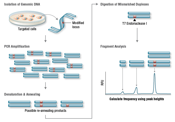
A widely used method to identify mutations is the T7 Endonuclease I mutation detection assay (36, 37) (Figure 3). This assay detects heteroduplex DNA that results from the annealing of a DNA strand, including desired mutations, with a wildtype DNA strand (37).
T7 Endonuclease I Targeting Efficiency Assay


Genomic DNA is amplified with primers bracketing the modified locus. PCR products are then denatured and re-annealed yielding 3 possible structures. Duplexes containing a mismatch are digested by T7 Endonuclease I. The DNA is then electrophoretically separated and fragment analysis is used to calculate targeting efficiency.
Another important parameter is the incidence of off-target mutations. Such mutations are likely to appear in sites that have differences of only a few nucleotides compared to the original sequence, as long as they are adjacent to a PAM sequence. This occurs as Cas9 can tolerate up to 5 base mismatches within the protospacer region (36) or a single base difference in the PAM sequence (38). Off-target mutations are generally more difficult to detect, requiring whole-genome sequencing to rule them out completely.
Recent improvements to the CRISPR system for reducing off-target mutations have been made through the use of truncated gRNA (truncated within the crRNA-derived sequence) or by adding two extra guanine (G) nucleotides to the 5´ end (28, 37). Another way researchers have attempted to minimize off-target effects is with the use of “paired nickases” (20). This strategy uses D10A Cas9 and two sgRNAs complementary to the adjacent area on opposite strands of the target site (Figure 2B). While this induces DSBs in the target DNA, it is expected to create only single nicks in off-target locations and, therefore, result in minimal off-target mutations.
By leveraging computation to reduce off-target mutations, several groups have developed webbased tools to facilitate the identification of potential CRISPR target sites and assess their potential for off-target cleavage. Examples include the CRISPR Design Tool (38) and the ZiFiT Targeter, Version 4.2 (39, 40).

Applications as a Genome-editing and Genome Targeting Tool

Following its initial demonstration in 2012 (9), the CRISPR/Cas9 system has been widely adopted. This has already been successfully used to target important genes in many cell lines and organisms, including human (34), bacteria (41), zebrafish (32), C. elegans (42), plants (34), Xenopus tropicalis (43), yeast (44), Drosophila (45), monkeys (46), rabbits (47), pigs (42), rats (48) and mice (49). Several groups have now taken advantage of this method to introduce single point mutations (deletions or insertions) in a particular target gene, via a single gRNA (14, 21, 29). Using a pair of gRNA-directed Cas9 nucleases instead, it is also possible to induce large deletions or genomic rearrangements, such as inversions or translocations (50). A recent exciting development is the use of the dCas9 version of the CRISPR/Cas9 system to target protein domains for transcriptional regulation (26, 51, 52), epigenetic modification (25), and microscopic visualization of specific genome loci (27).
The CRISPR/Cas9 system requires only the redesign of the crRNA to change target specificity. This contrasts with other genome editing tools, including zinc finger and TALENs, where redesign of the protein-DNA interface is required. Furthermore, CRISPR/Cas9 enables rapid genome-wide interrogation of gene function by generating large gRNA libraries (51, 53) for genomic screening.

The future of CRISPR/Cas9

The rapid progress in developing Cas9 into a set of tools for cell and molecular biology research has been remarkable, likely due to the simplicity, high efficiency and versatility of the system. Of the designer nuclease systems currently available for precision genome engineering, the CRISPR/Cas system is by far the most user friendly. It is now also clear that Cas9’s potential reaches beyond DNA cleavage, and its usefulness for genome locus-specific recruitment of proteins will likely only be limited by our imagination.

From NEB expressions Issue I, 2014
Article by Alex Reis, Ph.D., Bitesize Bio
Breton Hornblower, Ph.D., Brett Robb, Ph.D. and George Tzertzinis, Ph.D., New England
Biolabs, Inc.

Y- RNA

Y-RNA

Y RNAs have been found in all vertebrates in which they have been studied, as well as in C. elegans and the bacterium D. radiodurans. There are usually 1-4 Y RNAs present in a given species, and these Y RNA genes are usually found within a cluster on the same chromosome. They contain their own promoters and the distance between the genes as well as their order is well-conserved .
Studies surrounding the evolution of Y RNA genes indicate that a single ancestral Y RNA duplicated to give rise to two distinct Y RNA genes, which further gave rise to four Y RNA genes in the order Y5-Y4-Y3-Y1. The presence of less than 4 Y RNAs in some species suggests that some of these genes were lost through evolution .
Northern blot analysis of various vertebrate Y RNA samples probed with human Y cDNA showed the Y3 is the most conserved, Y1 is less conserved than Y3, and Y4 and Y5 are the least conserved.
significant partner RNAs or proteins: 
The RoRNP consists of two proteins that interact directly with Y RNA, Ro60 and La.  La contains an amino terminal RNA binding region (RNP-80) and carboxy terminus phosphorylation sites that modulate RNA binding ability. La binds to an oligouridine stretch found at the 3' end of Y RNA. Ro60 also contains an amino terminal RNA binding domain (RNP-80), and its overall conformation is also important for proper binding to Y RNA. Ro60 binds to the distal stem region of Y RNA at conserved nucleotides found within a bulged structure. Interaction of La and Ro60 with Y RNA have been proposed to protect and stabilize Y RNA, as well as aid in its translocation from the nucleus to the cytoplasm .  As a general mechanism, Y RNA binding to La and Ro may inhibit the chaperone activity of these proteins.
hY1 and hY3 have also been shown to interact more transiently with hnRNP K and I and nucleolin in the loop region. The significance of this is not as well studied, but may involve regulation of the chaperone activities of these RNPs and proteins .
Y RNA functionally interacts with components of the DNA replication machinery, such as RPA and PCNA.
mechanism of action: 
Studies revealing the structure of Ro60 suggest that Y RNA has the ability to inhibit Ro60's chaperone activity by binding to it . Ro60 binds misfolded RNAs via the external region of its N-terminal circular domain. 3' single stranded regions of bound RNA are threaded through the central cavity of this circle, and this is thought to be crucial for targeting misfolded RNAs for degradation or possibly for facilitating refolding. Y RNAs bind Ro60 at this same external region of the circular domain via their stem region. This sterically hinders the ability of misfolded RNAs to bind Ro60, implicating Y RNAs as a repressor of Ro60 activity. Similarly, regions in Ro60 required for its translocation to the nucleus seem to be masked by binding of Y RNA, and binding of Y RNA to hnRNP K and I has been shown to inhibit their splicing related chaperone activities , 
The mechanism for RoRNP mediated resistance to UV irradition in D. radiodurans and mouse ES cells is unknown.
The mechanism for Y RNA's role in DNA replication is unknown. It does not appear that Y RNA functions as a primer, and it does not involve interaction of Y RNA with Ro60.
cellular functions: 
Y RNAs are part of the RoRNP, which also contains the proteins Ro60 and La. Ro60 acts as a molecular chaperone that regulates proper folding and assembly of small, non-coding RNAs by binding them. For example, roles for Ro60 in regulating the maturation of U2 snRNAs, 23S rRNAs, and 5s rRNAs has been observed in mouse ES cells, D. radiodurans, and X. laevis, respectively . La also appears to have chaperone activities involved in regulating processing of RNAs and assembly of RNPs . Binding of Y RNA to Ro60 and La is thought to repress these chaperone activities . Similarly, hY1 and hY3 may inhibit chaperone activities of the more transient interacting partners, heterogeneous nuclear RNP (hnRNP) K and I . The most recent reports also suggest Y RNA regulates the localization of Ro60. While the functional roles of the different Y RNAs have not been elucidated, it has been proposed that their structural differences may confer the ability of the RoRNP to recognize different types of misfolded RNAs.
Studies in mammalian cells and D. radiodurans suggest that the Ro60 and Y RNA may also be important in resistance to UV irradiation. Increases in RoRNP particles (i.e. increases in levels of Ro and binding of Y RNA to Ro) in D. radiodurans and increases in the nuclear localization of RoRNPs in mouse embryonic stem cells occurs in response to UV irradiation, and inhibition either of these processes decreased survival, implicating a role for RoRNPs and Y RNAs in UV resistance, however the mechanism behind this is not known.
Y RNA has also been shown to have an essential role in DNA replication. Studies using isolated human nuclei in cell free DNA replication systems were used to show that hY1, hY3, hY4, or hY5 is required but not sufficient for replication in this system. This data also showed that there is redundancy among the hY RNAs in regulating DNA replication. These results were supported by in vitro data showing that siRNA knockdown of hY1 in HeLa cells decreased proliferation.

Molecular basis of vision revealed

Date:
September 30, 2019
Source:
Cornell University

Researchers have solved the three-dimensional structure of a protein complex involved in vertebrate vision at atomic resolution, a finding that has broad implications for our understanding of biological signaling processes and the design of over a third of the drugs on the market today.
The findings illuminate how signals from photons (particles of light) get amplified in the eye. More importantly, the study provides insights into how the largest family of cell membrane proteins -- G-protein-coupled receptors (GPCRs) -- work in humans.
"They're involved in almost all the biological processes in a human body -- how we perceive light, taste, smell, or how the heart rate is regulated or muscles contract -- and they are targets for over 30% of the drugs that are used today," said Yang Gao, co-first author of the paper and a postdoctoral researcher in the lab of Richard Cerione, the Goldwin Smith Professor of Chemistry and Chemical Biology and co-senior author.
There are over 800 GPCRs in humans that signal through about 20 different G proteins. GPCRs are responsible for sensing a wide range of outside signals -- such as hormones, light, and sense of smell and taste -- and inducing corresponding responses inside the cell. In vertebrate vision, the GPCR rhodopsin is capable of detecting the signal from just one photon and through the activation of the G protein transducin and downstream effectors, amplify it 100,000 times.
The researchers used cryo-electron microscopy to obtain atomic-resolution structures of the rhodopsin-transducin complex. The structures not only provide the molecular basis of vertebrate vision, but also reveal a previously unknown mechanism of how GPCRs in general activate G proteins.
"What we've learnt from these structures at an atomic level may be broadly applicable to other GPCR signaling systems," said co-first author Sekar Ramachandran, a senior research associate in Cerione's lab.
By learning more about how different receptors specifically couple with different G proteins, the researchers hope to gain insights into designing drugs that specifically regulate GPCR signaling. A lot of drug side effects occur when therapies are not specific enough and target both harmful and beneficial pathways, Yang said.
Hongli Hu, a postdoctoral researcher in Stanford's Department of Structural Biology, is a co-first author; Georgios Skiniotis, professor of molecular and cellular physiology and of structural biology at Stanford, is a co-senior author.

Story Source:
Materials provided by Cornell University. Original written by Krishna Ramanujan.

Careers in Veterinary Science


We human beings have dominated this world, but the world doesn't belong to human beings alone. There are many more species which not only help in maintaining the ecological and biological balance in the nature but are also useful to mankind in many ways. For example milk which is an important food all over the world, comes from animals. For meat and poultry, which again is consumed by a large number of people, has to come from animals and birds. Medicinal items and nutritional supplements are drawn both from natural and animal resources. We also keep animals as pet. A large population all over the world earns their living through farm animals including buffaloes, cows and goats. Though modern methods of cultivation have come, bullocks are still used for the purpose. All in all, animals very much belong to our world and we can't imagine our existence without them, in one way or the other.
Courses like MBBS, BDS etc. are much sought after by students completing their 10+2 with science subjects including biology. Such courses come under medical careers. People with these qualifications treat humans for different ailments. Animals and birds may also need treatment and there is a specialised field for that named as veterinary science. A person qualified in veterinary science is called veterinarian or vet (in short). The job of a veterinarian, basically, is to attend to the medical and healthcare needs of animals which include pets, livestock, zoo and laboratory animals. In cities and urban areas  the most common animals to be treated by them are dogs and cats. In others they may have to attend to cows, bullocks, goats, sheep etc. Different segment of veterinarians may be  involved in different requirements related to animals. Generally the job of a veterinarian consists of conducting physical examination, administering immunization, providing emergency and critical care, performing surgery and dental procedures and offering advice to people regarding care of their pets and livestock etc. Many of the animals maladies and infections may have adverse effect on people coming in the contact of such animals. So, veterinarians' role is also crucial to keep the  human society safe and healthy. By monitoring and protecting the health of animals that provide food for a large section of population, veterinarians ensure a safe food supply for them. If you are not averse to  animals, are comfortable in dealing with them and have  aptitude for medical science, you may plan to grow as a veterinarian.
Subjects of study
As a student of veterinary science you will be exposed to both theoretical and practical aspects related to animal health, anatomy, physiology, biochemistry, pharmacology, parasitology, toxicology, epidemiology, genetics & breeding, nutrition, gynaecology and obstetrics, radiology, surgery, clinical and farm practices, livestock products technology and production management, veterinary clinical practices, livestock farm practices etc. Largely the course is divided into basic and clinical sciences.
Requirements to become a veterinarian
Formal education to become a veterinarian can be pursued at graduation level. The nomenclature for this  graduation qualification is Bachelors in Veterinary Science, commonly known as B.V.Sc. In many cases the degree also includes the term animal husbandry.  The entry requirements are same as applicable for MBBS. The applicant should have completed H.S.C. (i.e. 10+2) with physics, chemistry and biology or biotechnology with English. Selection is usually based on performance in entrance examination conducted for the purpose.
Various states conduct this entrance test for admission to veterinary institutes in their jurisdiction. While most of the states have a separate examination for entry to B.V.Sc., few have a common/ combined entrance examination for this and other  professional courses  like B.Tech., B.Pharm.  etc.
Veterinary Council of India  conducts a national All India Pre Veterinary entrance examination/test. 15 percent seats in recognised institutes across the country are filled on the basis of qualifying scores in this examination. There are nearly 40 such institutes where this quota of 15 per cent is applicable.
Entrance examinations  are  generally conducted with a duration of  2 to 3 hours in which objective questions from physics, chemistry, botany and zoology  are asked.
The course duration for a degree in veterinary science is 5 and a half years of which one year is for intense internship.  A B.V.Sc. degree opens the door to work as  a vet. However many students prefer to join a  master's degree course, M.V.Sc. after graduation. The  course period for this postgraduate qualification  is 2 years, divided into 4 semesters and eligibility criteria is B.V.Sc. pass. To be eligible for M.V.Sc. , candidates have to appear in the qualifying test conducted by  Indian Council for Agricultural Research in collaboration with National Testing Agency.  Doctoral (Ph.D.) opportunities are also available for those interested in research. University Grants Commission norms apply here as in case of Ph.D. in other subjects.
Selecting  an Institute for study
Veterinary education is an old discipline of study offered at government universities and colleges since decades. Now  few private institutes have also started courses in the subject.
Indian Veterinary Research Institutes (deemed to be university) at Izat Nagar, Bareilly is  a premier establishment in veterinary education. However it offers course from postgraduation onwards only. Government has  established a number of animal sciences universities in the country as given below:
Bihar Animal Sciences University,Patna
Nanaji Deshmukh Pashu Chikitsa Vigyan Vishwavidyalaya, Jabalpur
Sri Venkateswara Veterinary University, Tirupati
Lala Lajpat Rai University of Veterinary & Animal Science, Hisar, Haryana
U P Pandit Deendayal Upadhyay Pashu Chikitsa Vigyan Vishwavidyalaya evam Gau Anusandhan Sanstha, Mathura
Tamilnadu Veterinary & Animal Science University, Chennai
Chattisgarh Kamdhenu Vishwavidyalaya, Durg
West Bengal University of Animal & Fisheries Science, Kolkata
Karnataka Veterinary, Fisheries & Animal Science University, Bidar
Guru Angad Dev Veterinary & Animal Science University, Ludhiana
Maharashtra Animal & Fisheries Science University, Nagpur
Kerala Veterinary & Animal Science University, Wayanad
Kamdhenu University, Gandhinagar
Madhya Pradesh Pashu Chikitsa Vigyan Vishwavidyalaya, Jabalpur
Rajasthan University of Veterinary & Animal Science, Bikaner
Prof Jayashankar Telangana State Agricultural Veterinary University, Hyderabad
Other than above there are a number of colleges/universities which offer graduate/postgraduate courses in veterinary science and animal husbandry, some of which are listed below.  where you may pursue a degree. The list is not in any particular order and is for  indicative purpose only:
Dr. Rajendra Prasad Agricultural University, Pusa
Birsa Agricultural University,  Kanke, Ranchi
Assam Agricultural University, Jorhat
Indira Gandhi Agricultural University, Raipur
Anand Agricultural University, Anand, Gujarat
Govind Ballabh Pant University of Agriculture & Technology, Pantnagar
Junagadh Agricultural University, Gujarat
P.V. Narsimha Rao Telangana Veterinary University, Hyderabad
Sardarkrushinagar Dantwada Agricultural University, Banskantha, Gujarat
Chaudhary Sarvan Kumar Himachal Pradesh Krishi Vishwavidyalaya, Palanpur
Swami Keshvanand Rajasthan Agricultural University, Bikaner
Narendra Dev University of Agriculture & Technology, Faizabad
Pondicheri University
Orissa University of Agriculture & Technology, Bhubaneshwar
Central Agricultural University ,Imphal
Jawahar Lal Nehru Krishi Vishwavidyalaya, Jabalpur
College of Veterinary Science  & Animal Husbandry, Salasih, Mizoram
Work opportunities
Supply of Veterinary professionals is limited so it may be relatively easier to find a job or settle down in career. This position  shouldn't be interpreted as lack of competition in job market .So you need to be better equipped. Those interested to work in government may join government veterinary hospitals at district and taluka levels which exist parallel to  government general hospitals/primary health centres. In many states District Livestock Development Officers (DLOs) are appointed  who in many cases are considered to be the incharge of Veterinary hospitals.
One good opportunity for which only a  graduate qualification is needed comes from public sector banks who allow candidates with B.V.Sc. to join as Agricultural Field Officer/Rural Development officer. Selection is based on  written test and interview. Private banks operating in rural areas and insurance companies also retain veterinarians.
Another option is to work with drug researchers and manufacturing companies. Poultry feed and pesticide companies also appoint veterinarians. Wildlife parks, zoos, sanctuaries, zoological parks etc. need the services of veterinarians on a regular basis. Most of such establishments are managed by government bodies. Bulk milk producers and cooperative organizations  need veterinary doctors.
Defence services and police departments  need services of veterinarians to look after dogs engaged for security and investigation purposes.
There is good business opportunity for Veterinary doctors to pursue private practice by establishing their own clinic. Alternatively you may join a big clinic and offer your services as a member in doctors team.
Those interested in business and marketing may take up M.B.A. in Agri-Business  after completing their B.V.Sc.
Teaching opportunities can be explored at the institutes listed above and others involved in Veterinary education.
Candidates with an aptitude for research may work as a research scientist at Indian Veterinary Research Institutes. Government of India has established a number of research establishments many of which conduct research on a particular animal. These organizations, as listed below, appoint  candidates with higher qualification in veterinary science :
National Research Centre on Camel, Bikaner
Sheep Research Centre, Mannavanur, Tamilnadu
Central Institute for Research on Goat, Makhdum, Uttar Pradesh
Central Sheep & Wool Research Avikanagar, Rajasthan
Buffalo Research Centre, Hisar
National Research Centre on Meat, Uppal, Hyderabad
Central Research Institute on Cattle, Hisar
National Research Centre on Yak, Dirang, Arunanchal Pradesh
National Research Centre on Equines, Hisar
National Research Centre on Mithun, Jhampani, Nagaland
There are few other research Institutes dealing with animals health  issues viz.
National Institute of Veterinary Epidemology, Bengaluru
National Institute of Animal Nutrition and Physiology, Bengaluru
CSIR-National Institute of High Security  Animal Diseases, Bhopal
Scholarship based research opportunities can be explored in foreign countries.
Challenges
Animals also communicate but differently from humans. They can't explain what ails them so it requires extra efforts to understand their pains and problems and then decide on a course of action.
Skills and traits for success
Veterinarians must show compassion towards  their animal patients and their owners. Willingness to learn, explore and to go the extra mile keep a veterinarian in good stead.
Useful websites to visit
If you want to further explore the field of veterinary and animal science, following websites of  related organizations  may provide you useful information:
Veterinary Council of India  https://aipvt. vci.nic.in
Indian Veterinary Research Institutes http://www.ivri.nic.in
Indian Council for Agricultural Research https://ntaicar.nic.in
National Testing Agency https://www. nta.ac.in

SOURCE- EMPLOYMENT NEWS, volume-28, 12-18, October, 2019